
OCTiX logo

Intraoral OCT.
Now Scan Inside and Out

Explore Lilivis solutions for scanning, design, milling, printing, and implant workflows.

Intraoral OCT. Now Scan Inside and Out OCTiX

FEATURE

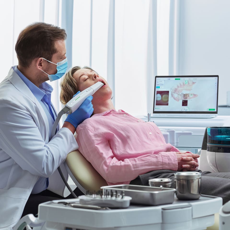
dentist-using-intraoral-scanner-on-patient

Revolutionizing Oral Scanning
with High-Precision OCT Technology

Huvitz's Optical Coherence Tomography (OCT) technology is an optical imaging technique that analyzes internal structures with high precision by irradiating light. By projecting infrared light, which is harmless to the human body, it provides high resolution below 60μm. This allows for high-resolution observation of the microstructure of teeth and soft tissues. The technology enables precise imaging of deep tissues in the teeth and gums, including dental caries (cavities), microcracks, periodontal diseases, and subgingival structures.

Currently, the Cone Beam Computed Tomography (CBCT) technology used in dental applications employs X-rays and provides a resolution level of 80μm–400μm. However, CBCT has limitations in observing microstructures such as fine cracks due to its low resolution for soft tissues (e.g., gingiva and pulp). Additionally, repeated imaging poses radiation exposure risks, raising patient safety concerns.

Huvitz's OCT technology offers a non-invasive diagnostic experience, free from skin incisions or radiation exposure, ensuring the safest and most comfortable diagnostic process for patients.

Certificate icon

Patent: TOMOGRAPHY CONVERGENCE-TYPE ORAL SCANNER

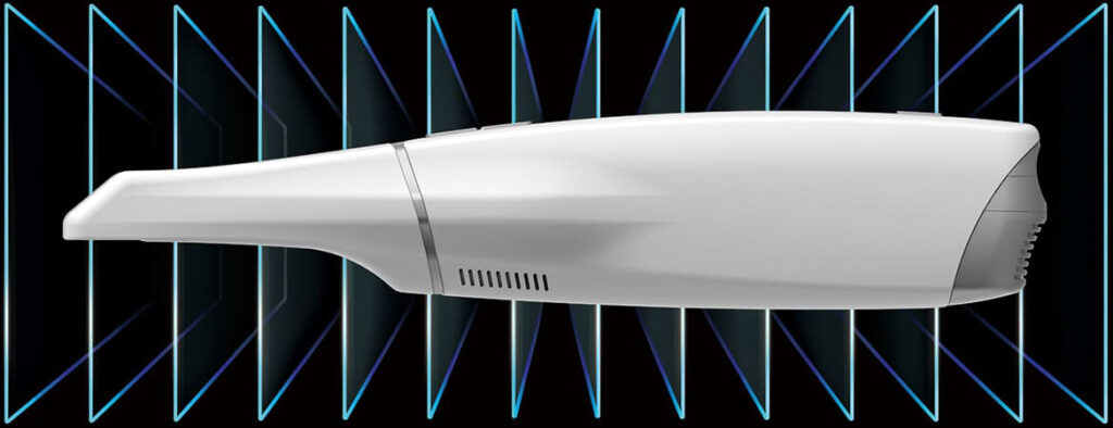
The "Tomography Convergence-Type Oral Scanner" patent describes a technology that simultaneously scans the external surface and internal structure of teeth to generate a comprehensive 3D image. By integrating Optical Coherence Tomography (OCT) scanning with intraoral scanning functions, this technology enables precise and extensive dental diagnostics. The data obtained from both scanning processes are combined through a dichroic mirror, allowing simultaneous observation of the external and internal structures of the teeth. This technology supports real-time 3D analysis, providing fast and accurate diagnostic results immediately after scanning.


[Patent No. 10-2458985, 12011336]

Enhanced Accuracy and Clarity with OCT icon

Enhanced Accuracy and Clarity with OCT

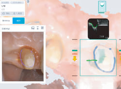
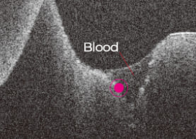
The Optical Coherence Tomography (OCT) interferometry imaging function penetrates gingival epithelial tissue without being affected by saliva or blood to accurately capture subgingival margins area. Experience a difference in technology with precise margin data unlike anything you've encountered before.

Simplifying Prosthesis Fabrication with AI Technology icon

Simplifying Prosthesis Fabrication with AI Technology

Real-time scan images integrated automatically with margin data detected by AI streamline the process. This integration facilitates the creation of optimal prostheses through a simple verification step, followed by processing with L-CAD software, eliminating complex editing.

Faster Scanning with a Wider and Deeper Area icon

Faster Scanning with a Wider and Deeper Area

Designed with a wide 15x18mm Field of View (FOV) and a maximum 22mm Depth of Field (DOF), the scanner enables broader area coverage with a single movement. This ensures a rapid and smooth impression acquisition process.
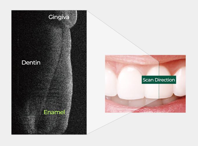
Veneer mode

Cross-sectional image
of a tooth by OCTIX

cross-sectional-image-of-a-tooth-by-octix

Veneer Scan
Process

Pre-operation

Pre-operation

After preparation

veneer-scan-process​

Enamel thickness

Sub-gingival finish line

Evaluating veneers

· Analyze enamel thickness and prosthetic adhesion.

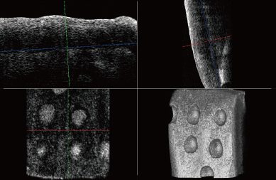
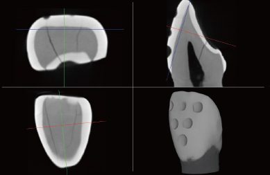
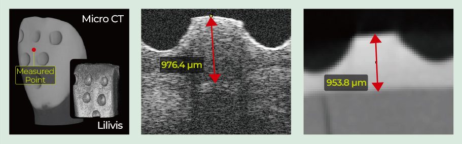
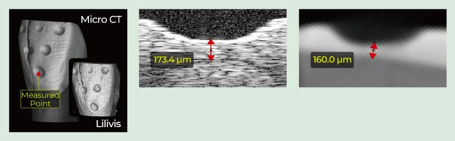
Accuracy of measured enamel layer

OCTIX

accuracy-of-measured enamel-layer-octix-fit

Micro CT

accuracy-of-measured enamel-layer-micro-ct-fit

Micro CT

accuracy-of-measured enamel-layer-micro-ct-fit

Before Prep.

3D

OCTIX

Micro CT

accuracy-of-measured enamel-layer-after-fit

After Prep.

3D

OCTIX

Micro CT

accuracy-of-measured enamel-layer-before-fit

Before Preparation deviation(OCT - Micro CT) : 22.6 μm
After Preparation deviation(OCT - Micro CT) : 13.4 μm

· High-resolution imaging at micrometer scale, exceeding Micro CT.

· Safe, repeatable imaging suitable for pregnant patients.

※This study was conducted Jointly by Seoul National University Dental Hospital and Huvitz.

Sub-gingival margin mode

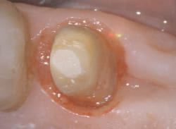
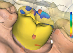
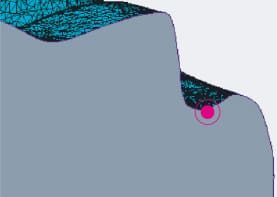
Real Margin Point Contact

real-margin-point-contact-sub-gingival-prep
Sub-gingival Prep
real-margin-point-contact-ios-scan
IOS Scan
real-margin-point-contact-design
Design(CAD)
real-margin-point-contact-octix
OCTiX(Accurate finish line)

OCTiX Process(Without Cord)

Another company

octix-process-without-cord-another-company-1
octix-process-without-cord-another-company-2

The Margin Point is on the surface

OCTIX

octix-process-without-cord-octix-1
octix-process-without-cord-octix-2

The Margin Point is under the surface

· Distinguishes gum and tooth for precise margin lines

· No need for cord insertion to measure margins.

TABLE FOR OCTiX

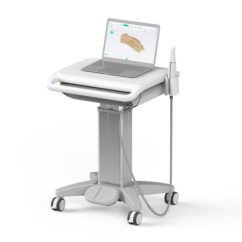
intraoral-scanner-cart-dental-3d-imaging-system

You can easily install the instruments and adjust their height with a simple pedal operation. The elegant white and silver color scheme blends seamlessly into various spaces.

DIMENSION 580 x 590 x 784.5 mm
WEIGHT 30kg
POWER supply AC 220V-230V, 50/60Hz
Travel Length 250mm
Speed 22mm/sec
Load Max 10kg
Others Up, down movement operation pedal included
Specification and design are subject to change without notice.

VIDEO

SUPPORT

If you need more product information, please download the data.

TECHNICAL DATA

Model​

LSO-100

Handpiece​

Dimensions : 277 x 40 x 57 mm   Weight : 270 g

Scanning

FOV : Up to 15×18 mm   DOF : Up to 22 mm

Imaging(OCT)

FOV : 6 mm  
Image Depth : Up to 5 mm

TIP

End Cross Section : 21.7 × 20.9 mm  Sterilization : Autoclavable (150 times per tip)

Main Body

Dimensions : 490 × 363 × 103 mm   Weight : 10 kg

Connectivity

Cable Length : 1.8 m   ​

* Distinguishes gum and tooth for precise margin lines

INQUIRE

Contact us Form

Service & Support

If you need support, please fill out your inquiry.
We will guide you through the next steps as quickly as possible.

Inquiry

Please fill out and send your inquiry
We will kindly guide you